
As Munk and Verhagen showed, a significant proportion of patients develop chronic symptoms after an ankle ligament injury:
- giving way
- repeated sprains
- pain
- stiffness
- locking
- swelling
The amount of disability form such symptoms has not been studied in detail. As many ankle injuries occur during sport, difficulty in returning to sport is a common complaint.
Functional and mechanical instability
It might be assumed that persistent giving way and re-injury of an ankle is because the ligaments, having been torn, no longer contribute to stability and therefore there is excess movement in abnormal directions. In fact, many patients believe this and expect that they need the ligaments repaired. However, not all patients with chronic ankle symptoms have abnormal ankle movement, even if their main symptom is giving way.
Freeman (1965) introduced the terms functional and mechanical instability. Functional instability is the complaint of giving way of the ankle; some have also used the term for a feeling that the ankle is about to give way. Mechanical instability is abnormal movement of the talus within the ankle mortise, usually demonstrated on stress radiography. Not all patients whose ankles give way (who have functional instability) have mechanical instability, and not all patients who have mechanical instability have functional instability.
The definition of mechanical instability varies from paper to paper. The most precise investigation was that of Karlsson et al (1991b). They measured anterior draw and tilt radiographs (see below for definitions) in 200 normal controls without ankle problems, 63 patients with bilateral functional instability and 57 with unilateral functional instability, using a stress jig. Their criteria for mechanical instability were chosen for maximum discrimination between the functionally stable and unstable groups: 10mm or more of anterior draw and/or 9deg or more of talar tilt. Where a contralateral functionally stable ankle was available for comparison, criteria of 3mm excess anterior draw or 3deg excess talar tilt were advised. Karlsson et al also documented the interobserver variability in measurement. However, few other authors have found such a close relationship between functional and mechanical instability.
Other causes of chronicity
Many factors, other than mechanical instability, have been advanced to account for chronic ankle instability:
- deficient proprioception
- peroneal weakness
- nerve injury
- central balance deficit
- altered reflex activity
- biomechanical factors
- generalised joint laxity
In addition, the associated injuries listed above may produce pain, altered function and even loose bodes in the ankle. There is evidence to support the presence of all the above factors, but not enough data to determine which factors are most important.
The main biomechanical factors are
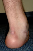
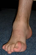
This patient presented with recurrent giving way of his ankle. He was not aware there was anything unusual about the shape of his feet!
hindfoot alignment : a varus hindfoot will tend to impose a varus/internal rotation force on the ankle, and in addition tends to limit the amount of compensatory movement available in the subtalar and midtarsal joints. Thirty percent of patients with cavovarus feet have ankle instability (Barrie 2000). Strauss (2007) identified hindfoot varus in 8% of patients undergoing primary ankle stabilisation but in 28% undergoing revision stabilisation. Vienne (2007) reported eight patients with failed ankle stailisation due to unrecognised hindfoot varus. Some cause of hindfoot varus, such as Charcot-Marie-Tooth disease, are also associated with peroneal weakness. A valgus hindfoot may place additional stress on the deltoid ligament and is associated with chronic medial ankle instability (Hintermann 2005).
tight Achilles tendon : limited ankle dorsiflexion means that the ATFL has to stabilise the ankle through more of the gait cycle and places more stress on a weakened ligament.