
Anatomy
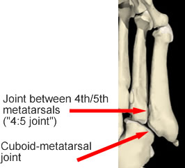
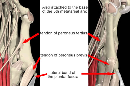
The fifth metatarsal forms the mobile lateral foot border. It has a broad base, expanded laterally to form the tuberosity, a narrow shaft and a fairly small head. The base articulates with the cuboid and with the fourth metatarsal. The anatomy of these joints and their ligaments has not been described in detail.
Biomechanics
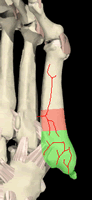
- the maximal stress occurs 3.4-4cm from the tuberosity (the zone of stress fractures)
- the maximal stress acting on the fifth metatarsal is 50% of the failure stress, supporting the view that fractures in this area are indeed stress or fatigue fractures.
Vertullo et al (2004) demonstrated torsional strain in the proximal fifth metatarsal under simulated axial loading and tendon contraction. They suggested this is due to the eccentric placement of the tendon insertions.
Orendurff (2009) studied the differences between the pressures under the 5th MT head and base during various sports manoevres as an indirect way of estimating bending forces. The largese pressure differences were during acceleration and running straight. Jump takeoff and landing and cutting right or left had low differences.
Raikin (2008) studied the hindfoot alignment in 21 patients with Jones fractures. Standing hindfoot alignment radiographs showed 18/21 to be in varus, and this was apparent clinically in 16.
