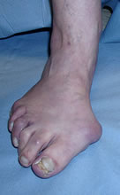
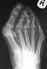
Introduction
Rheumatoid Arthritis (RA) is the most common form of inflammatory arthritis and affects about 1% of the world population – women three times more often than men. Early signs of RA include symmetric swelling of small joints of the hands or feet, which is associated with stiffness. Diagnostic criteria (developed by the American Rheumatism Association) include morning stiffness, swelling, nodules, positive laboratory tests and radiographic findings.
The foot and ankle are common sites of pathology. The inflammation can affect joints, tendons and ligaments. Vainio (1956) described foot disease in nearly 90% of rheumatology inpatients (848 of 955 feet). In a systematic review, Jaakola and Mann (2004) found foot and ankle symptoms in 90% of patients with RA.
Michelson et al (1994) found that 79% of university outpatients had ankle disease and 67% MTPJ disease; in about half of the patients these sites were the main problem. Twenty-seven percent had midfoot disease but only 5% considered it their worst problem. Interestingly, only 4% had accommodative shoe wear.
A number of studies of early RA have shown that the foot is one of the earliest sites affected, with 15% of first MTPJs affected within one year and 28% within 3 years (van der Heijde 1992). Calisir (2007), using MRI, found abnormalities in 70% of feet of newly diagnosed RA patients. Bouysset (1992) found that 50% of RA patients had a flexible flatfoot within 5 years of disease onset, but by 9 years the same proportion had stiff flatfoot deformities. Recent interest has focused on the use of MRI to help make the diagnosis of RA. Several studies have confirmed that the hands and feet are the most reliable sites in detecting early suspected disease (Boutry et al 2003).
RA patients with foot disease are more disabled, have poorer quality of life and possibly more active general disease (Wickman 2004). Foot and ankle surgery in RA is more likely to be complicated by infection than surgery at most other sites (relative risk 3.2 - den Broeder 2007).
A few rheumatoid patients develop enthesopathy with pain in the attachments of the plantar fascia and tendons (especially the Achilles tendon). However, this is much commoner in seronegative arthropathies such as ankylosing spondylitis, psoriatic arthropathy and Reiter’s syndrome.
Rheumatoid patients are challenging to treat, because of:
- Multiple joint disease with
- Significant general disability
- Interaction of disease sites such as the valgus knee and flatfoot (Keenan 1991)
- Effects on rehabilitation – upper limb disease makes it difficult to use crutches after foot and ankle reconstruction
- Systemic effects of rheumatoid disease, such as:
- Osteopaenia, which has negative implications on bone fixation
- Vasculitis leading to acute ischaemia, ulcers and wound breakdown
- Friable skin and subcutaneous tissues, compromising tissue handling and wound healing
- Neuropathy leading to pain and ulceration over stiff deformities - Wilson et al (2006) found that 60% of RA patients had reduced sensation in their feet compared with 12.5% of controls (mean age of both groups in 6th decade)
- Immune suppression with reduced resistance to infection
- Anaemia of chronic disease
- Pulmonary disease
- Complications of medical and other management
- Psychological and social effects of a severe, progressive, disabling disease in often young people
It is important to work closely with other members of a team including:
- Rheumatologist
- Physiotherapist
- Podiatrist
- Orthotist
- Nurse
- Orthopaedic specialists in other sites such as hip, knee and upper limb
- Radiologist
Clinical assessment
Physical examination of the rheumatoid foot and ankle should include a thorough assessment of the entire patient and observation of gait. Focal examination of the foot and ankle requires a systematic review of the skin, neurovascular status, range of motion, strength, overall alignment and assessment of deformities. Peripheral pulses must be carefully examined to assure adequate perfusion in potential surgical candidates. A vascular consultation should be obtained if there are any signs of arterial insufficiency which may affect surgical intervention.
Non-surgical treatment - general principles
Treatment goals in RA include:
- Control of synovitis and pain
- Maintenance joint function
- Prevention of deformities
A multidisciplinary approach is essential, involving pharmacotherapy, physiotherapy and occasionally surgery. Pharmacotherapy is the cornerstone of treatment in RA and has significantly reduced the number of patients requiring surgery in the past decade.
- Anti-inflammatory medication (steroids as well as NSAIDs)
- Disease-modifying antirheumatic drugs (DMARDs), i.e. Methotrexate, Hydroxychloroquine, Gold, Penicillamine.
- Immune-modifying drugs (AKA “biologics”)
There is limited data regarding the risk of postoperative infection in patients taking these drugs. Historically the it was usually recommended to stop anti-rheumatic medication in the perioperative period, to minimize the risk of infection and wound healing problems. However, recent studies did not show any evidence for this approach. Bibbo et al (2003) analysed 103 patients retrospectively and found no difference in the rate of wound healing problems and infections postoperatively with the use of NSAIDs, corticosteroids, hydroxychloroquine, methotrexate or gold. In a subsequent prospective trial, Bibbo et al (2004) showed a significant higher rate of complications in patients not receiving ant-TNFa agents. Den Broeder (2007) found no increased infection risk in patients who inadvertently continued biological agents through the perioperative period. Another prospective study of 388 RA patients (Grennan 2001) demonstrated that discontinuation of methotrexate therapy in the perioperative period was associated with higher infection rates.
Non-operative management of RA in the foot includes
- Physiotherapy
- Exercises can increase comfort and minimise disability in RA patients.
- A meta-analysis of the effectiveness of exercise therapy (Gaudin 2008) demonstrated improvements in muscle strength and aerobic capacity.
- Neuberger (2007) showed that the overall symptoms of fatigue, pain and depression in RA were improved by vigorous exercise
- Munneke (2004) found significantly less radiologic damage to the joints of the foot with high intensity weightbearing exercises than routine exercises
- Accommodative shoes (extrawidth/ extradepth)
- Padding over prominences
- Orthotics (custom made inserts or AFOs)
Surgery should be reserved for patients where
- Non-operative management has failed
- The deformity has become “unbracable”
The ankle
Michelson’s study shows that ankle disease is commoner than is often realised. The ankle should be examined in every patient with rheumatoid forefoot disease.
Ankle disease usually presents with pain and swelling in the ankle joint line. The joint is painful to move. Although the ligaments are eroded the ankle is not usually unstable. Some patients with severe erosion of the ankle may also have hindfoot valgus, although this is usually due to a combination of abnormal limb biomechanics, subtalar/talonavicular erosion and tibialis posterior tendonopathy (see below). The proximal limb joints, overall limb alignment and the forefoot should also be assessed.
Plain radiographs will demonstrate joint damage, erosions and periarticular osteopenia – standing AP/lateral are standard views. If there is hindfoot valgus a standing hindfoot alignment view (Saltzmann and el-Khoury 1994) helps to define the contributions of ankle and subtalar tilt.
Non-surgical treatment
Steroid injections are often helpful on an empirical basis, although there is no good evidence base for their use and in other joints they may not be much better than placebo. Rest/immobilisation and custom orthotics/bracing are often beneficial. An AFO might be required to improve alignment of the ankle and hindfoot.
Surgery
Synovectomy
A long-term Japanese study reported recurrent disease in only 10% of ankles at 10-20 year follow-up of open synovectomy (Akagi et al 1997); however, other studies suggest that improvement is maintained for only 4 years, similar to radiation synovectomy (Nakamura et al 2004; van der Zant 2004).
Arthrodesis
Until recently this was the only real option for end-stage ankle arthritis of whatever aetiology. The traditional British method was that of Charnley with a destructive anterior transverse approach and external fixation with a semi-stable frame. This had a high incidence of infection and non-union. Arthroscopic fusion has deveoped to the point where it is now the standard techniue for most foot and ankle surgeons.
Arthroscopic ankle fusion is normally performed with the patient supine using anterior portals. The technique of forming the portals is described in the section on arthroscopy.
Open fusion can be done using anterior, posterior and lateral approaches. A short medial arthrotomy is sometimes a useful adjunct to a lateral approach, although an extensile medial approach can be used to access talonavicular, subtalar and ankle joints. The lateral approach exposes the lateral malleolus, which can be excised and morcellised for graft, or removed, decorticated and attached as a large graft at the end. An alternative technique for graft harvest is to use an acetabular reamer to remove the lateral malleolus. The superficial peroneal and sural nerves, and their communicating branches, are at risk.
The anterior approach is the same as for ankle replacement, between tibialis anterior and EHL and exposing back to both malleoli. The deep peroneal/dorsalis pedis bundle is at risk, and the medial branch of the superficial peroneal nerve can cross the incision.
In arthroscopic fusion, synovitis and spurs are cleared to expose the joint line. Curettes and burrs are then used to remove the remaining articular cartilage and freshen the subchondral bone so that multiple bleeding points are seen. It is important to clear all the way to the back, usung the FHL tendon, usually seen through the posterior ankle capsule, as a landmark. It is also important to clear the gutters, the malleoli and the medial and lateral surfaces of the talus and to ensure that no osteophytes on the talus prevent compression of the talus into the mortise. Once preparation is complete, fixation is usually with two or three cancellous lag screws inserted over guidewires. Intra-operative check Xrays often show a gap between the joint surfaces, but as long as it is is not too big and fixation is secure, the gap will disappear as healing progresses.
Many series of open ankle fusion have used flat cuts with a power saw or osteotome to excise the joint. Others use techniques to increase stability and congruency, such as anatomical resection of the joint surfaces in the manner of arthroscopic fusion, or a chevron cut (Kopp 2004). Preservation of the malleoli increases stability.
However the joint surfaces are prepared, the ankle should be positioned in neutral plamtar/dorsiflexion, 5 deg valgus and 5-10 deg external rotation (Buck 1987). Fixation in most series was with 2-4 cancellous bone screws. The stability of the fusion can be increased with an anterior or lateral plate, blade plate or ring fixator.
The ankle is normally protected with a backslab immediately after surgery and this is converted to a cast when the wounds are satisfactory. Where the construct is stable enough, partial to full weightbearing can begin quite early; we allow patients with arthroscopic fusions to bear weight to comfort at 2 weeks. There are a variety of regimes for reducing splintage, but no clear evidence of what regime is best to optimise fusion rates without spending unnecessary time in cast.
Results
Arthroscopic fusion
Over 15 series of arthroscopic ankle fusions have been reported. The largest series is that of Winson (2005). 118 fusions were reported, of which 105 were followed clinically for a mean of 65m. 109/118 procedures fused at a mean of 12weeks. Most non-unions happened early in the series, and subsequently the authors immobilised all patients for a minimum of 12 weeks. Smoking was twice as common in non-unions. There were three superficial and one deep infection, one malunion and two pulmonary emboli. Winson recommended arthroscopic fusion even if the ankle was in valgus or varus provided the forefoot was plantigrade.
Gougiolias (2007), however, carried out arthroscopic fusions even if there was mortise deformity and the foot was not plantigrade, by excising bone from the mortise arthroscopically to align the ankle. 30/78 patients had a pre-operative coronal deformity of >15deg, but none had more than 3deg post-operatively. Five patients had simultaneous arthroscopic subtalar fusion. There were 5 delayed unions and 2 non-unions; time to union was 2months extra in smokers. There was one PE, one nerve injury and 6 patients had subtalar pain.
Overall about 550 arthroscopic fusions have been reported with a total fusion rate of 93.3%. Several series have commented on the quicker rate of union in arthroscopic procedures although this can be influenced by the intervals at which radiographs are taken, and all series diagnosed union on plain radiography rather than CT. Myerson (1996) noted a mean time of fusion of 8.7 weeks in arthroscopic versus 14.5 weeks in open fusions, while Nielsen (2008) noted that 90% of arthroscopic and 57% of open fusions were united at 12 weeks.
The arthroscopic technique is now the standard method of fusing the ankle unless there is severe deformity, bone loss or revision surgery. However, it requires advanced arthroscopic skills. It is feasible in patients whose soft tissues would not be suitable for open surgery. However, there has never been a RCT comparing open and arthroscopic fusion.
Open fusion
A study of patients 20 years after ankle fusion (Fuchs et al 2003) reported reduced SF-36 pain, physical functioning and emotional disturbance scores, and moderate Olerud ankle scores. Most patients wore customised footwear, but few had walking aids. All but one had returned to work. There was significant progressive OA in the other hindfoot joints. The surgery included more external fixation than would be expected in a current population, and most patients had what would now be considered mal-unions. Another study (Buchner et al 2003) reported little or no pain and restriction of activity in 92% of patients at an average of 9.3 years follow-up.
An independent study of fusion in OA (Anderson et al 2002) found that the true fusion rate was 80-89% but the clinical and radiological outcomes were not the same. In rheumatoid disease, Felix et al (1998) reported union in 96% of 26 ankles at 2-8 year follow-up, and no pain. Although many studies are quite old and use techniques that are uncommon now, the risk of non-union is almost certainly higher in open than arthroscopic fusion.
Revision fusion
Cheng (2003) reported 18 revisions, 10 of which were for non-union, 7 for mal-union and one for infection. Screw fixation was used in all but one case and 17/18 fused including the infected case, taking a mean of 4.8months. At a mean follow-up of 40months the mean AOFAS score was 71/100 (range 38-86). Easley (2008) reported 45 patients including 8 infected cases. 11 had screw fixation, 12 had a tibiotalocalcaneal fusion with a nail and 22 with bone loss or infection had a ring fixator. At a mean 50months follow-up, 40/45 were fused, 5 of whom had required revisions. There were 5 amputations and 4 malunions and most complications were in the patients who had the ring fixators.
Total ankle replacement
Replacement of the ankle has been around for over 30 years. However, early prostheses did not reproduce the biomechanics of the ankle well and had a very high failure rate. Second-generation prostheses from the late 1980s onward introduced improved engineering, often with three components. 10-20 year results are now being published (Kofoed 2004, Buechel et al 2004), indicating success in over 90% of patients. However, these are the series of the designers of the various prostheses, and further results from the “real world” may not be quite so optimistic. The Wrightington series (Wood and Deakin 2003) is a realistic and critical account with 5-8 year follow-up and 92% 5-year success. This has now been updated (Wood 2008) with 80.3% survival at 10 years.
Results in OA and RA are similar (Kofoed 2004, 1998; Wood and Deakin 2003). Ankle prostheses also seem to be best uncemented; the long term results of the STAR ankle have been significantly better in the uncemented design (Kofoed 2004). Like all joint replacements, failure occurs and may require revision replacement or fusion. The failure rate for revision arthroplasty is high and most patients will be better revised to a tibiotalocalcaneal fusion.
Fusion versus ankle replacement
It is only appropriate to compare fusion and replacement for patients without major deformity, infection, bone loss or neuropathy – these would only be candidates for fusion. However, there have been no RCTs comparing replacement and fusion in patients in whom both would be an option. In addition, outcome measures in single-procedure series are so varied that it is difficult to compare them.
The only comparative study is that of Saltzmann (2009, 2010). This was an FDA approval study in which patients were assigned to STAR ankle replacement or open ankle fusion through a fibular osteotomy with screw fixation, according to the surgeon doing the procedure. There was also an open series of ankle replacements. The main outcome measure was the Buechel-Pappas ankle score. This improved by a mean of 40 pints for the STAR group and 26 points for the fusions, mainly accounted for by significant improvements in most of the functionss cores. However, there was no significant difference in pain relief or patient satisfaction. There were more intra-operative problems, nerve injuries and wound problems in the STAR group, but similar infection rates of 4.4% in the replacements and 7.6% in the fusions (all but one minor). The union rate in the fusions was 64/66 and there appear to have been 7/415 loose replacements at 24 months (this is not easy to work out), with one revision for wear and 4 mobile bearing fractures. At an average follow-up of 4 years the clinical results of TAR were similar to those of fusion (Saltzman 2010). However, the arthroplasty group showed better pain relief and more postoperative complications that required surgery.
A systematic review of the literature by Haddad et al (2007) found the overall reported results of both procedures to be similar, with about 70% satisfactory results after both procedures. The overall non-union rate for ankle fusion was 10% and the arthroplasty survival rate 77% at 10 years. There was more variation in results for arthroplasty.
Clinical series of ankle replacement
Gougoulias (2009) published a systematic review of 13 series amounting to 1105 ankle replacements, of which the Agility, STAR and Hintegra were commonest. Only 4 studies had more than 10 years’ follow-up. Survivorship rates were variable, satisfaction rates varied from 79-97% but 23-60% of patients still had some pain. The Swedish arthroplasty register figures (Henricson 2007) found a 5-year survival of 78%, but this improved to 86% after the first 3 cases. The Nowegian register (Fevang 2007) reported 5-year survival of 89% and 10-year of 76% in mostly STAR ankles. The Finnish register (Skytta 2010) reported a 95% 5-year survival in a 50% rheumatoid population using mostly AES and STAR ankles.
Wood (2009) reported the only RCT in ankle replacement, comparing 100 STAR ankles with 100 Buechel-Pappas ankles. The survival rates at 6 years were 955 for the STAR and 79% for the BP (the difference was not significant) In both groups survivorship was lower with increasing coronal plane deformity.
These studies tend to highlight need for revision as the main outcome measure. At 12 years, the survivorship of the current cementless Buechel-Pappas implant was 92% (Buechel et al 2004), but clinical results were presented only as excellent 88%, good 5%, poor 7% (75 patients). Twelve-year survival for the STAR ankle was 95%, with a mean Kofoed ankle score of 91.7/100. The long-term survival rate is based on small numbers in each study. The Wrightington series (Wood 2008), reported 200 ankles followed for a minimum of 5years and a mean of 7.3 years. The 5-year survival was 93.3% and the 10-year survival 80.3%. The mean AOFAS hindfoot score for pain improved from 0/40 pre-operatively to 35 at final follow-up and the functional score from 28/60 to 40. Complications and the learning curve are detailed. As in the RCT with the BP ankle, survivorship was significantly lower in the presence of coronal plane deformity, and Wood recommends 15deg valgus or varus as the maximum for successful ankle replacement. However, Hobson (2009) reported 123 STAR ankles including 32 with coronal plane deformity of 10-30deg, mostly varus. Survival at 5 years was 86% for the neutral ankles and 75% for the deformed ankles, which was not a significant difference. Nine patients in the deformed group needed additional corrections – 6 calcaneal osteotomies and 3 lateral ligament reconstructions.
None of these studies comment on OA in adjacent joints.
Kinematics and gait
As might be expected, ankle fusion affects the range of motion and kinematics more than does replacement, and certain designs are closer to normal than others (Valderrabano et al 2004a,b). Gait analysis is much closer to normal in replaced than in fused ankles (Butcher 2004, Piriou 2008). Hence it is plausible that ankle replacements would place less stress on other joints, both in the tarsus and proximally. Nevertheless, this requires clinical confirmation.
Naal (2009) found that 2/3 of 101 patients were active in sports 4 years after ankle replacements. The commonest sports were swimming, cycling, fitness training and downhill skiing and the patients participated for an average of 4.4hr/week. 35% had radiolucencies around the tibial implant but this did not affect the likelihood of sports activity.
There is not enough data to indicate whether fusion or replacement is to be preferred for patients in whom either procedure would be an option. At about 10 years clinical success rates appear similar. Ankle replacement probably has a small functional advantage but failure rates vary widely and there is a much higher rate of secondary surgery.
It is possible that the onset of OA in other joints reduces the success of fusion thereafter. The improved kinematics of ankle replacement probably reduce the wear on adjacent joints, and this could be important, particularly in patients with multiple joint pathology. The improved range of movement is anecdotally useful to patients, but there are no comparative studies.
Hindfoot disease and flatfoot deformity
Rheumatoid disease of the hindfoot and midfoot joints was fairly common in Michelson’s series, although most patients found the ankle or forefoot more of a problem. Pain and swelling in the sinus tarsi is usually due to subtalar synovitis. Disease in the other tarsal joints usually presents with local pain and swelling.
Many rheumatoid patients develop a flatfoot deformity – 50% at 5 years in Bouysset’s (1992) series had a flexible deformity, but by 9 years tarsal joint destruction had developed and the deformity became rigid. The cause of the flexible deformity has been attributed to:
- Abnormal proximal limb mechanics. Keenan (1991) carried out a thorough study of rheumatoid patients with and without flat feet involving clinical examination, gait analysis and electromyography. She concluded that the main causes of the deformity were muscle weakness, adaptive gait changes due to pain and knee valgus.
- Joint laxity due to rheumatoid disruption of ligamentous attachments and stretching of the capsule. Keenan considered that the gait anomalies she identified acted on weakened joints to produce the deformity
- Tibialis posterior insufficiency (TPI). By analogy with the non-rheumatoid adult acquired flatfoot, weakness of a damaged tibialis posterior has been investigated. Jernberg (1999) found 80% of rheumatoid flatfoot patients could not do a single foot tiptoe test and 20% had weakness on strength testing. However, MR showed tendonopathy in both flatfoot and control patients equally. Several studies have failed to find tibialis posterior ruptures on ultrasound or MRI and are often quoted as evidence against TPI as a cause of the rheumatoid flatfoot. However, Coakley (1994) and Masterson (1995) found few ruptures but most tendons had tenosynovitis or tendonopathy; it is now recognised that this may be enough to provoke TPI without an actual rupture. Only Keenan found little clinical evidence of TPI in her flatfoot patients.
It seems likely that the deformity is due to a combination of these factors.
Patients with hindfoot valgus may do less well after forefoot surgery. Stockley (1990) found abnormal first ray loading and increased pain after Kates-Kessel-Kay procedures in patients with valgus hindfeet.
Non-surgical treatment
As at other sites, early disease can be managed medically or with local steroid injections. Clinical assessment of affected joint correlates relatively poorly with MR, and so MR imaging may be useful in deciding which joints to inject.
The evidence on the use of orthotics is inconclusive, with effectiveness varying from study to study. A systematic review by Clark (2006) found that although methodolgy is generally poor, there is evidence that casted orthotics can improve pain in the valgus foot, and can prevent deformity progression. Corrective orthotics can, of course, only be useful in patients with mobile deformities. In fixed deformities, accommodative orthotics may still be useful in improving comfort and preventing skin breakdown.
Surgery
Popelka (2010) reported 26 isolated talonavicular fusions in patients with RA, talonavicular joint destruction and tibailis posterior insufficiency. At a mean follow-up of 4.5y, mean AOFAS midfoot score improved from 48.2 to 88.6/100. There was one non-union. Eight patients continued to have pain in the foot, two severe. Joint disease progressed in one subtalar and one calcaneocuboid joint. This series implies that isolated fusion can work even in patients with RA, although there are some uncertainties in the resporting of this series that make generalisation difficult.
Most patients who require surgery for intractable pain or deformity will have disease in several hindfoot joints and require triple fusions (unlike patients with osteoarthritis, where disease affecting one joint predominantly is common and single joint fusion generally to be preferred). Figgie et al (1993) reported 40 triple arthrodeses in RA, with 5% non-union. Eight patients united in >10deg valgus, of whom 5 had ankle pain and 3 had undergone ankle fusion. Knupp (2008) reported 24 triple fusions in 20 patients with RA. All had flatfoot deformity. There were no non-unions or malunions, but 8 patients had superficial wound infections. Arthritis had progressed in 15 midfeet and 10 ankles, but this did not affect outcome. The mean VAS pain score was 47/100, AOFAS midfoot score 70/100 and short musculoskeletal function instrument 45 for dysfunction and 38 for bother. SF-36 scores were just below normal values.
Flatfoot deformity may require quite extensive joint resections and medial column shortening (Henderson et al 2002). Stabilisation with compression screws or staples produce similar reported rates of union and clinical benefit. A technique of removing and rotating a dowel of bone, without stabilisation, has been described but produces a high symptomatic non-union rate.
Forefoot
Assessment
In early RA the MTP joints are commonly affected. The prevalence of forefoot deformities in patients with chronic RA has been reported to reach 90%. MTP joint inflammation leads to capsular distension and eventually to loss of soft-tissue constrains. Weight-bearing in the presence of instability, articular cartilage destruction, and subchondral bone resorption results in the common rheumatoid forefoot deformities:
- hallux valgus
- clawing of the lesser toes
- subluxation/ dislocation of the lesser MTP joints
Some patients present with early flexible or semi-flexible deformities (type 1 or 2). However, the majority have severe clawing with fixed subluxation or dislocation of the MTP joints when referred for surgery. The toes, dislocated onto the dorsum of the metatarsal heads, no longer share load bearing in late stance phase. The plantar plate of the MTP joint is also dislocated onto the top of the head and locks it down by the “plunger effect” (Stainsby 1997). The dislocated toe and plantar plate draw the plantar fat pad forward by their connections to the plantar fascia, leaving the metatarsal heads exposed in the sole. Hence these patients often complain of a sensation of “walking on pebbles”. This is discussed further in the page on lesser toe deformities.
There is some controversy about the relationship between the altered forefoot mechanics induced by the flatfoot deformity and the development of hallux valgus. The abnormal laxity induced in all the first ray joints, especially the 1st MTP joint, is probably most important in creating the valgus deformity.
Occasionally referral is precipitated by the development of an ulcer over a stiff deformity, usually the medial prominence of the 1st MT head, under a lesser MT head or over a PIP joint. Ulceration may be due to peripheral neuropathy.
A few patients present with stiff painful 1st MTP joints without deformity, more like hallux rigidus than valgus.
Assessment of a patient with rheumatoid forefoot problems should always include a review of other joints, the overall limb alignment and examination of the joints and alignment of the ankle and hindfoot. Patients with hindfoot valgus do less well after forefoot reconstruction than those with normal hindfeet (Stockley 1990).
Check skin integrity and look for neuropathy and vasculitis.
Look under the forefoot for exposed metatarsal heads. Check the reducibility of toe deformities. If the MTP joint is reducible, how unstable is it? (draw test) Are the tender areas or calluses over the PIP joints dorsally or at the tips of plantar-flexed toes? If toe deformity is mild and most of the pain comes from the MTP joints, feel for synovitis – if in doubt an ultrasound can be helpful.
Non-surgical treatment
The first line of management of patients with rheumatoid forefoot disease is usually accommodative shoes with enough space to remove pressure on stiff deformities, and insoles to reduce pressure on the metatarsal heads. Clark (2006) found that both hard and soft insoles can improve forefoot pain, and can prevent hallux valgus progression. One study found that patients preferred metatarsal dome to metatarsal bar orthotics. Patients with severe deformities or peripheral neuropathy may need full moulded total contact insoles.
Ulcers will usually heal with pressure relief as for diabetic ulcers, even if there is neuropathy. Occasionally early surgical debridement will be necessary for osteomyelitis related to an ulcer. Once healed, the foot can be protected with accommodative shoes and moulded insoles. Sometimes ulcers with break down again unless the pressure is relieved by surgical reconstruction, and the risk of this may be considered so high that early surgery is recommended.
Surgery
Patients with pain and pressure problems which cannot be relieved by accommodation and orthoses may be offered surgical reconstruction. General control of systemic disease and, particularly, of vasculitis, should be established before surgery and ulcers should, if possible, be healed.
Management of the first MTP joint
First MTP joint without erosive disease
Mulcahy (2003) and Hulse both reported series in which some patients had excision of the lesser MT heads with no surgery on apparently normal 1st MTP joints. At 3 years Mulcahy found that 25% of the first rays had required surgery, and in Hulse’s series the rate was 46% at 6.5y.
Thordarson (2002) reported 11 patients with disease in the lesser MTP joints and symptomatic hallux valgus without apparent synovitis or joint erosion. A variety of joint-preserving procedures, mostly chevron osteotomies, were performed in association with lesser MT head excisions. By 3 years 11 had developed synovitis and/or joint destruction of whom 5 had undergone fusion, and 3 also required revision of their lesser ray surgery. On the other hand, Nagashima et al (2007) reported 80% satisfactory results 5y after Hohman osteotomy to the hallux and (probably) Helal-type osteotomies ot the lesser rays, and Berg et al (2007) reported satisfactory results at a minimum of 6y after scarf osteotomy to the hallux and Weil osteotomies to the lesser rays. Only Berg discussed disease severity – three of their patients had progressive disease and required fusions, and all had severe joint disease at presentation. It is also possible that preserving the metatarsal heads improved Nagashima and Berg’s results.
We are prepared to leave asymptomatic 1st MTP joints untreated but warn patients that there is a risk of at least 25% that they will have problems within 5 years. If there is hallux valgus but a good joint we are prepared to offer corrective surgery as for a non-RA deformity but warn patients that they may develop progressive disease and require a fusion. Alternatively we would be prepared to offer a fusion as primary treatment.
End-stage first MTP joint
The options are:
- Excision arthroplasty of the base of the proximal phalanx (Keller)
- Excision arthroplasty of the head of the first metatarsal (Mayo)
- 1st MTPJ fusion
- 1st MTPJ replacement, with or without 1st MT osteotomy to correct metatarsus primus varus
Excision arthroplasty has been most extensively reported. However, Mann and Thompson reported good results after fusion of the first MTP joint in association with excision of lesser MT heads, and considered that this gave more stability and fewer lateral calluses. Coughlin (2000) promoted this view in a very detailed paper. Other series, however (eg Beauchamp 1984, Hughes et al 1991, Mulcahy 2003) reported that although patients with fusion often had fewer central calluses and better gait parameters, the overall clinical results were much the same.
There is, unusually, one RCT in this group – Grondal et al randomised 31 patients to 1st MTP fusion with screw fixation or Mayo-type excision arthroplasty in association with leaser MT head excision. At 5-7y follow-up the fusion patients had fewer calluses, fewer shoe adaptations and slightly better centre of pressure position on pedobarography. However, there was no difference in the foot function index, satisfaction, insole use or hallux alignment.
Vandeputte et al reported a retrospective comparison of Keller-type excision arthroplasty with meticulous soft tissue reconstruction versus 1st MTP fusion, combined with lesser MT head excision. The Keller group had more recurrent hallux valgus and less 1st ray loading, but fewer calluses, and the overall clinical results were similar.
Only retrospective cohort studies are available for 1st MTP replacement. Hanyu et al (2001) reported 10-year follow-up on 39 silastic arthoplasties with lesser MT shortening. Although over half had implant subsidence and 21% synovitis, only 3 patients were dissatisfied. Shi (2007) reported 15 silastic arthroplasties with proximal 1st metatarsal corrective osteotomy and (mostly) lesser metatarsal shortening. There was only one non-progressive lucency around the implants and the AOFAS forefoot score averaged 82.
A case could be made for any of these first ray procedures and provided patients are appropriately counselled it probably does not matter which is used. There is considerable need for further RCTs.
Management of subluxed/dislocated lesser MTP joints
The options are:
- Excision of the metatarsal head (Hoffman, Fowler, Kates-Kessel-Kay – the differences are mainly about surgical approach)
- Excision of the base of the proximal phalanx (Newman/Fitton, Stainsby – taking most of the phalanx)
- Combined excision arthroplasty (Clayton)
- Open reduction of the MTPJ and metatarsal shortening (Helal, Weil)
The goal is to achieve enough bony shortening to reduce the toe, and this can be achieved by any of the above procedures. If metatarsal heads are excised, all should be removed as new problems tend to develop under retained heads. Indeed, the commonest complication of Hoffman-type procedures is recurrent painful callus formation under metatarsal stumps that have either been left long or have partially re-formed (10-40%). To avoid this most authors recommend care to produce a smoothly-curved parabola.
Stainsby (1997, 2001) designed his procedure around the plantar fascia, which is the main plantar flexor of the normal toe and also controls the position of the metatarsal fat pad. Sufficient phalanx is excised to allow toe reduction, but the metatarsals are not resected so that the bone and soft tissue lengths will be the same. Ironically, Mann and Thompson (1992) described all the important features of the Stainsby operation without, apparently considering the plantar fascia function – then proceeded to excise the metatarsal heads. Newman and Fitton (1983) described an operation in some ways similar to Stainsby, but clearly without the plantar fascia concept.
Metatarsal shortening osteotomies are designed to reduce the toe but preserve stability. They are inapplicable in severely eroded MTP joints (Hanyu 2001, though without clear data). A few series (Hanyu 2001, Shi 2007, Berg 2007, Niki 2010) have reported shortening osteotomies in RA with apparently good results, although without detailed reporting of the durability of the lesser ray correction.
There are no RCTs comparing lesser MTP procedures in RA. Reported results from cohort series are comparable, although MT head excision has a significant revision rate. There are many more reports of MT head excision than any of the other procedures. Probably any of these procedures could be justified with appropriate patient counselling.
Surgical approaches
Most older papers describe a transverse plantar approach for lesser metatarsal head excision, giving access directly to the prominent metatarsal heads. Often the 1st metatarsal head was also excised through this incision, although sometimes a medial or dorsomedial approach was used. Fowler (1959) added the excision of an ellipse of skin to draw the fat pad down, and this has been repeated by other authors eg Kates et al (1967). As Stainsby (1997) and Briggs (2001) showed, excision of a skin ellipse in unnecessary – the fat pad is controlled by its enclosing plantar fascia and will reduce with the plantar plate.
Concern about wound healing with a plantar approach, and the need to avoid early weightbearing, led to the use of dorsal approaches. A transverse dorsal approach gives excellent access to all MTP joints. Alternatively, longitudinal approaches on the first ray and 2nd and 4th intermetatarsal spaces also allow surgery on each MTPJ. For phalangectomies, incisions running into the toes are required. Briggs and Stainsby (2001) designed curved incisions in each to after studies on digital skin perfusion, and with careful soft tissue handling these heal with few problems.
In fact, plantar incisions generally heal with few problems. Barton (1973) reported 38% wound problems, but most series report 10% or fewer; indeed, van Loon (1992) had more wound healing problems (though fewer calluses) with dorsal incisions. It has been suggested that multiple dorsal incisions make it more difficult to resect metatarsal heads in a smooth arc, but Coughlin (2000) achieved a good arc in 45/47 feet. Patients who have plantar incisions are usually advised to avoid forefoot weightbearing in the early post-operative phase (eg Karambande 2007), which may interfere with rehabilitation in patients with multiple joint problems and poor balance.
The East Lancs Foot Service procedure of choice is a 1st MTP fusion with Stainsby procedure to the lesser rays. We do all lesser rays even if one or two are symptomatic or undeformed: these patients are likely to develop deformity in residual rays over the succeeding years. If the patient has previously had a Hoffman-type procedure we may offer the same on the other side if required. A failed Hoffman usually requires reduction of one or two stumps to make a smooth arc, and sometimes reduction of plantar plates from the dorsal surface of MT stumps.