
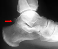
Os trigonum in plantigrade position
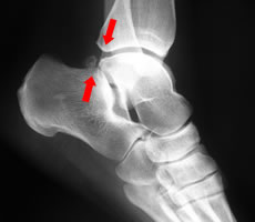
In plantar flexion the os trigonum is "nut-crackered" between the calcaneum and the posterior tibial lip
If there is clinical evidence of an inflammatory arthropathy this should be evaluated with the appropriate blood tests: FBC, ESR and rheumatoid factor tests are normally sufficient; in a possible spondyloarthropathy the HLA B-27 may be added.
Plain films will show spurs and loss of joint space. A lateral film in plantarflexion may demonstrate impingement of the os trigonum or posterior talar process.
Both ultrasound and MR can identify synovitis, impingement and ligament tears. MR is useful to evaluate the FHL tendon, but fluid collections around this tendon are not unusual and may be due to synovial communication with the ankle.
However, impingement is largely a clinical diagnosis. In our practice imaging is mainly used for patients with intra-articular or atypical pain.