
Hallux valgus is a clinical diagnosis made by simple observation, and no investigations are required to confirm it. Investigations have two functions in hallux valgus:
- identify important underlying conditions such as rheumatoid arthritis, osteoarthritis or infection
- assess the severity of the deformity and thus judge what would be required to correct it
Hence most patients only need radiographs (or any other tests) if an operation is the only way to improve their symptoms, and they wish to have one. You may feel this point is being laboured, but patients often turn up in the clinic with X-rays (supine, hence of limited value) when they either do not need, or do not want, an operation.
Radiography
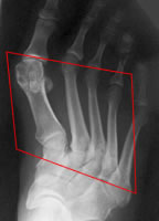
The main investigation for assessment or planning is a standing dorsoplantar radiograph of the foot. A standing lateral view adds some information on the alignment of the first ray and the quality of the 1st MTP joint. Occasionally, a sesamoid view adds useful information about the state of the sesamoid-metatarsal joint. Oblique views are rarely useful, as are other imaging modalities.
Non-weightbearing views tend to underestimate the severity of hallux valgus, and may give misleading information about alignment and relative metatarsal length - they should not be used routinely.
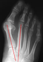
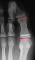
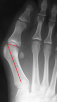
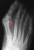
The dorsoplantar radiograph gives information about:
Severity of deformity
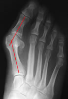
Hallux valgus angle
Intermetatarsal angle
Hallux interphalangeus angle
Distal metatarsal articular angle
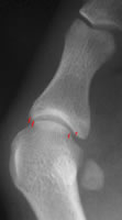
MTPJ congruency
Sesamoid position
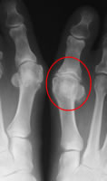
MTPJ arthritis
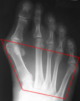
Forefoot shape: L splayfoot, R metatarsus adductus